I’d like to share with you an interesting case of a 3.6 with six canals.
In addition to the pleasure you can get from a free demonstration of the phases and procedures of such a retreatment, I think I can use this case to make some considerations.
I have always thought of myself as a good endodontist, ever since my degree. At the beginning I used to work with the naked eye, occasionally using, with low satisfaction, 3.5x goggles with no coaxial light that slightly improved my view of the endodontic field.
However, the fact that I often found the MB2 canal in the upper sixth was the best proof for my belief that magnification was not necessary and that I was a bit of a “chosen one” of endodontics, one of the few who can find the fourth canal with my eyes closed.
And, as a matter of fact, comparing how I worked back at the time and how I work now, I was basically working with my eyes closed!?
After thousands of canals and having started to always use goggles with coaxial light first and the microscope after, I’ve understood a few things.
First of all, as a new graduate I was indeed better than the majority of dentists who don’t have any idea of how you can decently perform a devitalization, if only for my theoretical knowledge and the fact that I was always using the dam, the mechanical equipment and thermafils. But, anyways, I sucked.
Secondly, whoever says that it’s not necessary to use goggles first (“I don’t want to risk and become dependant on them”?) and then the microscope (“after all, I can do everything with my goggles!”) simply does not have the experience and the means to make these statements. And I say so because I did it too!
The third point I want to make here, is that if you know what you could or should look for and you have the tools/instruments to see what you are doing, then things are not that difficult anymore.
Endodontics surely require a certain manual dexterity, great precision and a lot of patience.
And it also requires having all the instruments that technology makes available to us. Nickel-titanium rotary instruments, apex locator, irrigating solutions, a device for warm condensation, etc.
I would add the microscope and a cone beam for the more complex cases.
But the key to becoming a great endodontist is KNOWING HOW TO IMAGINE
Yes, that’s it. You need to be able to visualize the exact strategy you are going to use to manage the case even before you open the teeth. Exactly as a pilot can visualize and run the racetrack, before the start line,  with his eyes closed.
You must predict all the obstacles you are going to face.
You need to be ready to look for exceptions, because teeth are not all the same!
In the past, I gave for granted that all incisors had one canal, except for some sporadic lower incisors where I could find, almost by chance, two canals. The lower premolars had only one canal, and that famous Weine’s class IV of the lower first premolar, occurring in the 25% of cases, was a useless bogeyman.
Lower molars had 3 canals, except for some first molars with two distal canals.
The upper first molar had almost always the fourth canal and that was my strong suit, the one that made me a great endodontist.
However, with time, I have learnt that the extra canals are there. And there are a lot of them. Even so, only few people know that they can be there, and even fewer people know how to find them.
You don’t believe it, do you? Just have a look at this very interesting page from Dr. Mario Venturi’s website: http://www.endodonziamauroventuri.it/Anatomia%20sistema%20canalare.htm. And that’s it, if I see an x-ray of a lower incisor now, I check straightaway if it’s likely to have two canals, which is a very frequent thing.
If an upper first molar in the preoperative x-ray has somehow unusual roots, it could have three canals.
If a lower first premolar has a canal that disappears at a certain point, it’s likely to be a case of Weine’s class IV.
50% of the lower first molars have a middle mesial canal. And sometimes that happens for lower second molars too.
The upper second molar can have two canals. Or also just one. And often it’s the preoperative x-ray that tells you what the case is.
The lower second molar can have a C-shaped canal.
Moreover, the lower first molar can have an independent radix entomolaris at distolingual level.
And I have learnt that many of these anatomies can be predicted if you understand how to read x-rays that, even if two-dimensional, become a three-dimensional image for me, an expert endodontist. This happens exactly because I have learnt to visualize what kind of three-dimensional shape can have impressed on the x-ray I’m examining.
Sure, you need experience.
However, no one has ever told me how to visualize particular anatomies and I am sure that, having a good teacher that shows you how to do it, it’s possible to learn faster than I did.
In the retreatments then, if you see decent treatments that failed, you need to learn to suppose what might have happened. And here you are, a first molar like the one in the video you’ll see in a while. You have a decent treatment, but there is a lingual fistula. Therefore, in this case, I start the retreatment already knowing that there will probably be a middle mesial canal. I don’t simply retrace the canals thinking that, just by disinfecting a bit better and placing the dam, the granuloma will disappear, because this will hardly ever happen!
Needless to say, in order to be able to anticipate what you will find, you need experience. But sometimes CBCT can help you. I’m not one of those who think chat it’s better to do a CBCT in all the endodontic cases in order to get as much informations as possible. At first it can be useful doing a CBCT when you intuit the presence of some exception from initial x-ray: in these cases
I’ll give you an example.
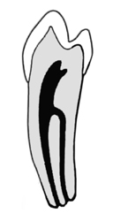
If I see a lower premolar that, in the x-rays, looks like it’s been well treated, but it has a big granuloma, I need to expect a vertical fracture or a second canal. The first times I looked for a second canal, I was always looking towards the vestibule aspect. Only after perforating a couple of premolars, I found out that the second canal, in the lower premolar, is always lingual. At the beginning you think it’s impossible, because looking at the teeth from occlusal it looks like you’re perforating it while you extend lingually. However, with CBCT and a vestibular perforation, you quickly understand how lower premolars with two canals are done and, at the end of the treatment, you will see that, opening the pulp chamber in the right way, these canals are well centred in the tooth, even though it seemed impossible before. And you understand it even better if you imagine you’re opening a tooth like this.
Do you understand, then, why you cannot find this canal if you don’t know it exists and if you don’t know that, when present, it’s in that position? We will definitely talk about the lower premolars in other posts.
For now, I felt it was important to give you some piece of advice.
If you assume that you don’t treat many teeth and therefore you cannot be that unlucky and find a premolar with three canals, you will never find it. But you will experience a lot of failures.
Actually, you will never experience any failures and you will remain convinced of this because your patients will have gone to some other dentist.
Only coming as little comfort to you and bad luck to you patient: whoever will come after you, won’t probably be any better than you. Unfortunately, nowadays, dentists still don’t know how to practice endodontics, although the instruments we have make it much easier for us than it was for Schilder and the fathers of modern endodontics.
My endodontics professor at the university asked to be paid for each canal and said that was the only way to get motivated to find them. I don’t know if that’s true. I find canals as a passion and because endodontics is fun if you know how to do stuff other people can’t do.
And, ironically, I reckon that canal treatments can fail now even more than when I just graduated. That’s because, back at the time, I didn’t do follow-up x-rays. And probably also because I didn’t notice some initial failures when my patients went to a different dentist.
As usual, I wrote too much. I’ll leave you to the video I promised I would post.
It’s a 3.6 with three distal and three mesial canals. The tooth had a fistula at lingual level on the mesial root. The disto-lingual and middle-distal canals were confluent, and I transformed them into one canal with a big apex and a favourable access that convinced me to obturate them with Biodentine. I obturated the other canals with Thermafil.
I hope I can transmit the emotion I feel when completing a case like this. Endodontics is beautiful if you know how to perform it. Enjoy the video and until next time!
Stefano
